From the branch of a lung to the crease between two toes, every twist of bodily tissue requires an immense system of chemical and physical processes to form.
Now Princeton University researchers have identified a third new process to probe developing tissue and organs — energy. In a study published April 22 in Science Advances, researchers describe how cellular metabolism in developing tissue helps determine the eventual shape of organs like lungs and hearts.
By taking precise measurements of a key molecule called ATP, which stores and releases the body’s energy, and mapping concentrations of ATP over the length of a tissue in a growing embryo, the team was able to know precisely where and when a fold would appear. This predictive power gives scientists a new lens on organ development and opens the door to engineering healthy tissue or addressing disorders with greater control.
“What the study tells us is that we need to be considering energy and metabolism as an intricate part of everything the cell and tissue does, including something that had seemed, previously, to be so far removed as the generation of force,” said principal investigator Celeste Nelson, the Wilke Family Professor in Bioengineering and professor of chemical and biological engineering.
Previous frameworks for studying organ development did not consider a cell’s energy budget as a relevant metric, according to the researchers.
“Before this, you usually would think about the energy budget of the whole embryo,” said Bezia Lemma, the paper’s first author. He said what did not exist is a clear picture of how energy changes vary across the space of individual organs.
The key innovation was to reimagine how metabolism acts on the structures of a developing body — in effect, to view metabolism in spatial terms at the level of both the cell and the tissue.
“Energy metabolism in this context, of cells doing things to build tissues, has mostly been ignored in the past,” Nelson said. “We lacked tools, we lacked insight. Metabolism was always seen as this kind of boring subject that you have to memorize, and then you move on.”
Lemma joined Nelson’s lab as a postdoctoral fellow with a background in physics and a specialty in understanding how energy drives motion at small scales. He wanted to build on Nelson’s previous work explaining tissues in terms of their physical forces and see if other patterns emerged based on cells’ underlying energy systems.
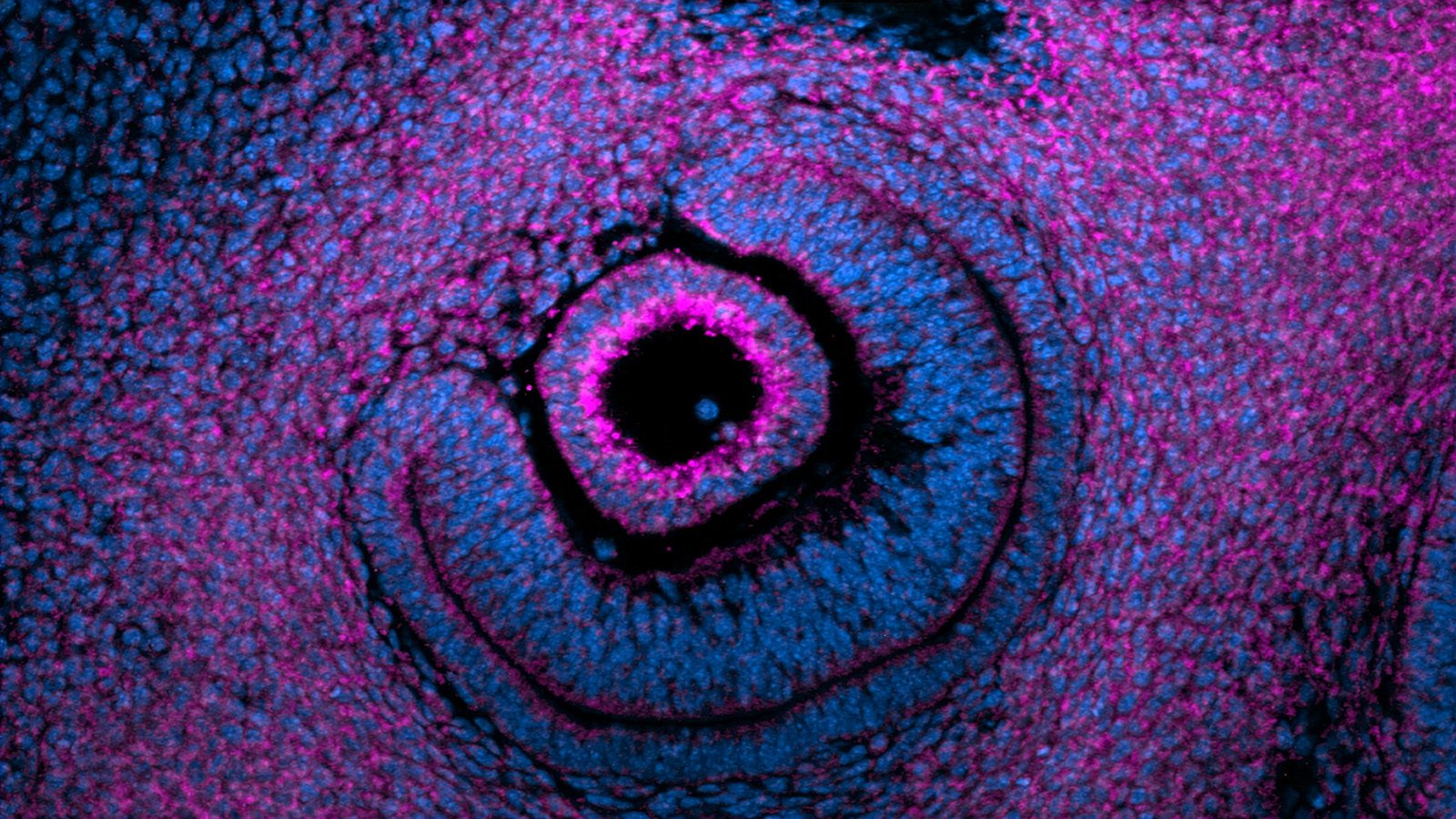
What he found surprised them both. Mitochondria, which release ATP and are often called the powerhouses of the cell, would crowd a particular point along one side of a developing tissue just before a crease appeared, the first fold of a developing nose or eye.
“The way the eyeball functions is related to its shape, right?” Nelson said. “If you don’t get that shape, you can’t see very well. And the source of age-related loss of vision is an alteration in the shape of the lens and the eye in general.”
Addressing shape using the predictive power of energy metabolism could unlock new technologies to probe and improve human health.
While the developing eyeball in mouse embryos stood out as the most striking organ they studied, the researchers looked at several other systems including fruit flies and chicken lungs, where they first identified the phenomenon. In all of the systems they studied, they observed similar spatial patterns, where an enormous amount of mitochondria appeared at a specific location, released ATP, and a fold would form.
“This is the era of spatial transcriptomics, where people are thinking about gene signaling in space,” Lemma said. “We’re trying to do the same with energy and forces.”
==
The paper, “Patterns of mitochondrial ATP predict tissue folding,” was published April 22, 2026 in the journal Science Advances. Research reported in this publication was supported by the Eunice Kennedy Shriver National Institute of Child Health and Human Development and the National Heart, Lung, and Blood Institute of the National Institutes of Health under grant numbers HD111539, HL164861, HD099030 and HL166311; and by the National Science Foundation under grant numbers 2134935 and NSF DMR-2011750. In addition to Lemma and Nelson, the authors include Megan Rothstein, Pengfei Zhang, Bridget Waas, Marcus Kilwein, Safiya Topiwala, Sherry X. Zhang, Anvitha Sudhakar, Katharine Goodwin, Elizabeth R. Gavis, Ricardo Mallarino, and Andrej Kosmrlj.