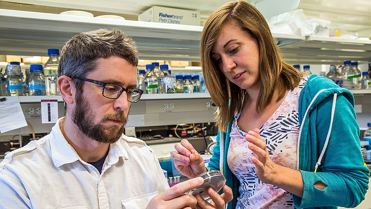
Researchers at Princeton University have harnessed AI to understand how drugs affect the dynamics of vital structures within the cell, introducing a tool that can map the shape of these structures to functional outcomes and shed light on important markers of health.
To do this, a team led by Cliff Brangwynne examined the shape changes of biomolecular condensates, tiny droplets in living cells that drive transcription and other gene regulation processes and have been linked to diseases including Alzheimer’s, ALS and cancer.
“The central problem in biology is how do you get emergent structure from individual molecular interactions,” said Brangwynne, the June K. Wu ’92 Professor of Chemical and Biological Engineering and principal investigator of the study. “The key innovation here was to develop a way to learn from the images and classify the patterns that are emergent.”
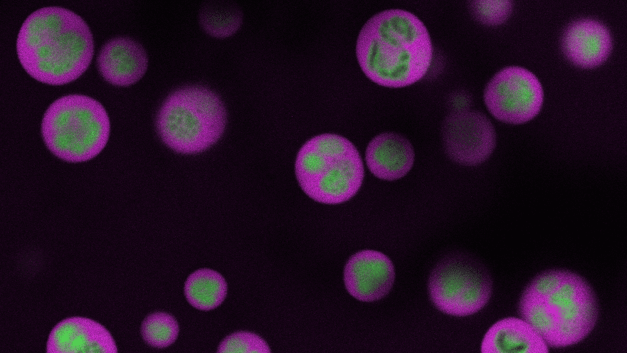
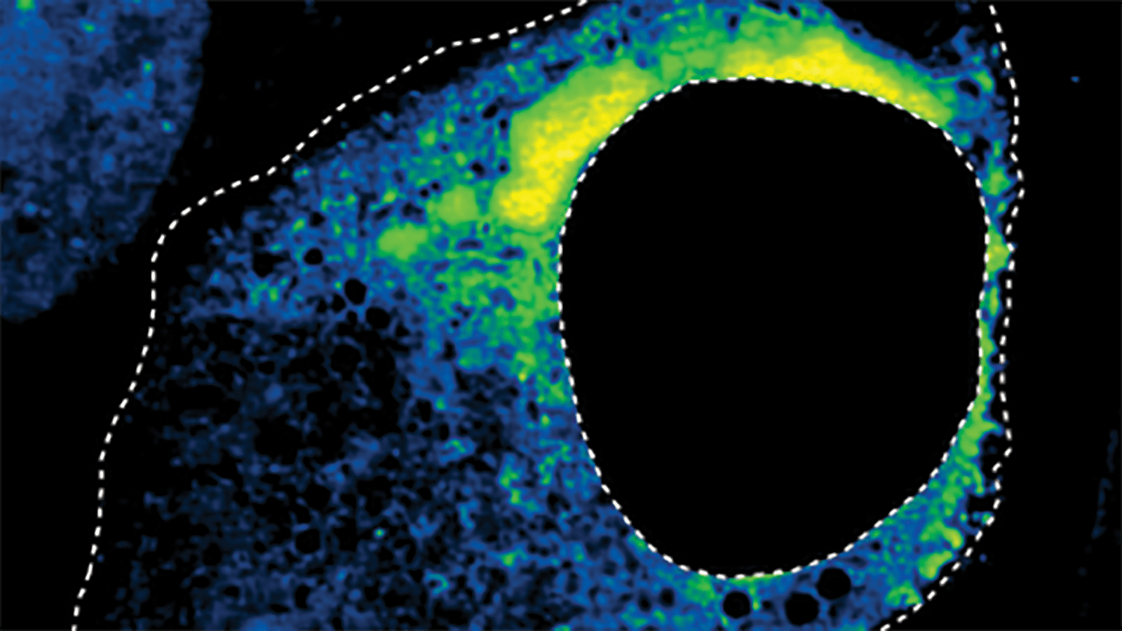
The study, published June 4 in the journal Cell, focused on a condensate called the nucleolus, responsible for assembling the tiny machines that build proteins. The team used an advanced microscope to image nucleolar shape changes in hundreds of human cells under a range of drug-controlled conditions. They then fed those images, difficult to interpret even for a highly trained scientist, through a machine-learning tool they had built for this purpose. The tool was able to sort the images into four basic categories based on the shape of the nucleolus — three categories that the researchers expected and a fourth that was unexpected, which the team later determined to be entirely new.
Anita Donlic, postdoctoral researcher, and Troy Comi, research software engineer, of Brangwynne’s lab, trained a neural network on tens of thousands of images of cells containing healthy, spherical nucleoli, plus two well-known atypical nucleolar forms: one shaped like a cap and another shaped like a beaded necklace, according to the researchers.
Members of the research team (left to right) Lifei Jiang, Lennard Wiesner, Anita Donlic, Cliff Brangwynne, Nima Jaberi-Lashkari. Photo by Wright Seneres
These cap and necklace shapes have been linked to a range of cellular stress responses, making them useful markers for testing the effects of drugs or gene therapies. For example, cap shapes can arise from a treatment known to disrupt processes that create the RNA needed to assemble the tiny protein-making machines. Necklace shapes can arise from a class of drugs that disrupt a separate RNA-related process.
After the initial training, the team ran a panel of drugs to see how each drug affected nucleolar formation. They used the neural network to visually measure changes in the condensate’s development, finding that different concentrations of drugs caused different degrees of change in both caps and necklaces.
The neural network found that two known anti-cancer drugs caused caps, a phenomenon not previously reported for those drugs, said Donlic, the paper’s first author. This suggests that these drugs may be affecting nucleolar function in ways that were not previously appreciated.
For a third drug, called topotecan, the network discovered a totally new nucleolus shape that the researchers labeled flower.

While topotecan was known to inhibit an enzyme used in DNA replication, Donlic showed that a loss of this enzyme — TOP1 — induced the flower shape and uncovered the enzyme’s role in maintaining nucleolar organization by regulating RNA processing.
“No one’s seen this flower morphology before,” said Brangwynne, who is also director of the Omenn-Darling Bioengineering Institute. “The network flagged it as not fitting neatly into the other three categories.”
These findings point the way toward a robust system for monitoring and evaluating cellular responses to drugs at a single-cell level.
The team also tested their neural network on other condensates related to RNA processes, observing similar dose-and-response results for drugs specific to nuclear speckles (a hub for messenger RNA activity) and condensates from respiratory syncytial virus.
This finding underscores the need to unlock these molecular-level mysteries that are easy for human analysis of basic factors like size and shape to overlook. “You could be missing other important features,” said Donlic. “Things that could tell you there’s new biology.”
The paper, “Deep Learning of Functional Perturbations from Condensate Morphology,” was published June 4, 2026 in the journal Cell. In addition to Brangwynne, Donlic and Comi, the authors include Sofia Quinodoz, Nima Jaberi-Lashkari, Krist Antunes Fernandez, Lifei Jiang, Lennard Wiesner and Ai Ing Lim of Princeton University. Support for the project was provided in part by the Howard Hughes Medical Institute, the Princeton Center for Complex Materials (NSF MRSEC DMR-2011750), the St. Jude Collaborative on Membraneless Organelles, the Chan Zuckerberg Initiative Exploratory Cell Network, and the Princeton Laboratory for Artificial Intelligence.