Princeton University researchers have developed a method aimed at improving how doctors implant medical devices in soft tissue, using the body’s natural adhesive properties to hold implanted materials in place.
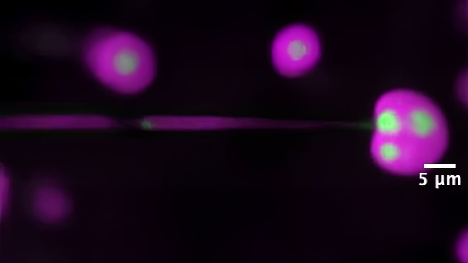
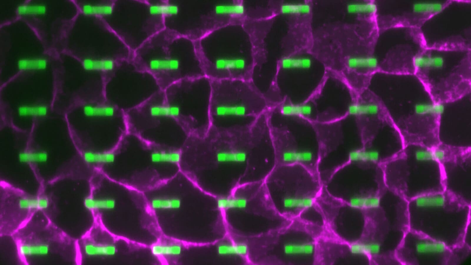
A team led by Daniel Cohen, associate professor of mechanical and aerospace engineering and the Omenn-Darling Bioengineering Institute, caused individual cells to wrap around an arch-shaped 3D plastic structure that is one quarter the size of the cells, or 25 times smaller than the width of a human hair. When cells wrapped around the arch, parts of their membrane met beneath the arch and created an embrace that locked the cells to the structure.
“We were asking, is there a way to use the machinery cells already have to recognize and hold on to each other — to hold onto the world around them?” said Cohen. “If you can find a tiny structure that can mimic the ‘secret handshake’ of the cell, you could make large arrays of these structures on implant surfaces and maybe make cells in soft tissues more stably attach to the implant.”
While integrating prosthetics into bone has become routine, implanting devices through soft tissues like skin, gums or the brain can still lead to infection, inflammation and the tissue pulling away from the implant. This is an important issue in applications such as medical screws, drug catheters and neural probes. Making soft-tissue cells more stable near the hard surfaces of a prosthetic is the key to lower rates of failure, the researchers said.
Their work was published in the journal Advanced Materials on May 24.
To address the problem, Cohen’s team turned to a protein called cadherin. Cadherins are what cells use to stick to other cells. Surprisingly, in cases where a cell wraps around an object, cadherins also enable that cell to stick to itself — “like shaking hands with yourself,” said Cohen. This prompted the team to explore making nanoscale 3D architecture to leverage the cells’ self-sticking qualities.
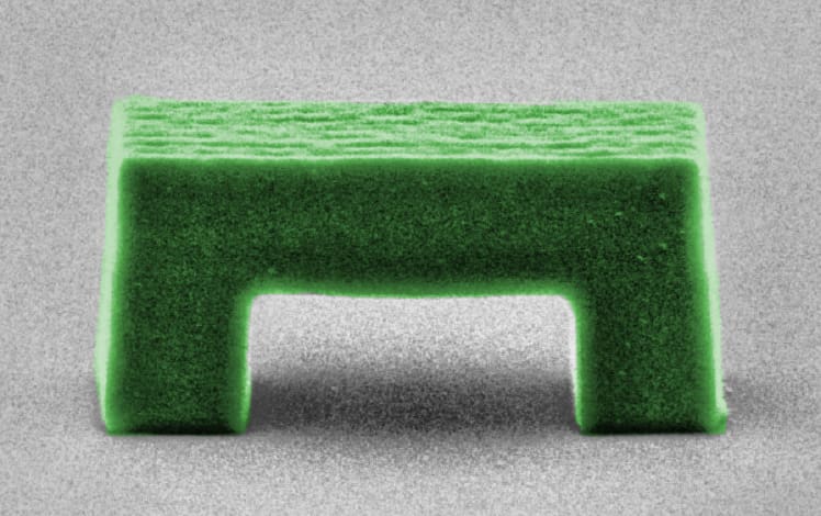
The first arch the team printed was about half the size of a cell. While this worked, it only caused cells to adhere strongly in about 60% of cases. To improve the system, the team 3D printed a range of nanostructures with different sizes and shapes. They found that smaller was better, with arches about a quarter of the size of a cell working well more than 80% of the time. Next, they found that sharpening the arch into a trapezoid improved the results further, allowing 93% of cells to stably attach to the structure.
Not only did this shape work in different types of cells — skin, kidney and stem cells — it also left the cells much more strongly attached. That meant the cells moved more slowly than in other cases, leading to a more stable overall system.
“That told me there was something special about this arch shape,” said Hannah Kim, a graduate student in Cohen’s lab and co-author on the paper.
The cells that formed self-adhesions around these arch-shaped structures lasted in that embrace for at least 24 hours, a key benchmark for creating a stable adhesion.
The team next plans to try this kind of 3D material coating with actual 3D tissue models such as engineered skin layers, more closely mimicking clinical uses.
The paper, “Engineering cellular self-adhesions inside 3D printed micro-arches to enhance cell:biomaterial attachment,” was published on May 24 in Advanced Materials. Co-authors included Anamika Singh, Lauren Rawson and Margaret Miao. Support for this project was provided by the National Institutes of Health and the National Science Foundation.