") Organized by the Imaging and Analysis Center at Princeton Institute for the Science and Technology of Materials (PRISM) and the scientific publisher, Nature, the conference, “Frontiers in Electron Microscopy for the Physical and Life Sciences” included more than 30 technical talks and panel discussions on emerging methods and the latest technologies. The 180 participants also had the opportunity to tour Princeton’s own cutting-edge microscopy facilities run by PRISM. The three-day conference received significant sponsorship from industry and academia, with lead support from Thermo Fisher Scientific and ExxonMobil.
Organized by the Imaging and Analysis Center at Princeton Institute for the Science and Technology of Materials (PRISM) and the scientific publisher, Nature, the conference, “Frontiers in Electron Microscopy for the Physical and Life Sciences” included more than 30 technical talks and panel discussions on emerging methods and the latest technologies. The 180 participants also had the opportunity to tour Princeton’s own cutting-edge microscopy facilities run by PRISM. The three-day conference received significant sponsorship from industry and academia, with lead support from Thermo Fisher Scientific and ExxonMobil.
“This conference is really unique in that it brings together scientists from both the physical and the life sciences to explore the new horizons of research now becoming available through electron microscopy,” said Dean for Research Pablo Debenedetti, the Class of 1950 Professor in Engineering and Applied Science and a Professor of chemical and biological engineering.
“We are excited to work with Nature to bring to Princeton the world’s leading scientists in this rapidly growing field, a field that offers the opportunity to probe biological and physical structures that are so tiny that they’ve been beyond our capability to explore,” said Debenedetti. “And yet these tiny structures are so essential to our health and to our technological capabilities. This is like being explorers in a new land.”
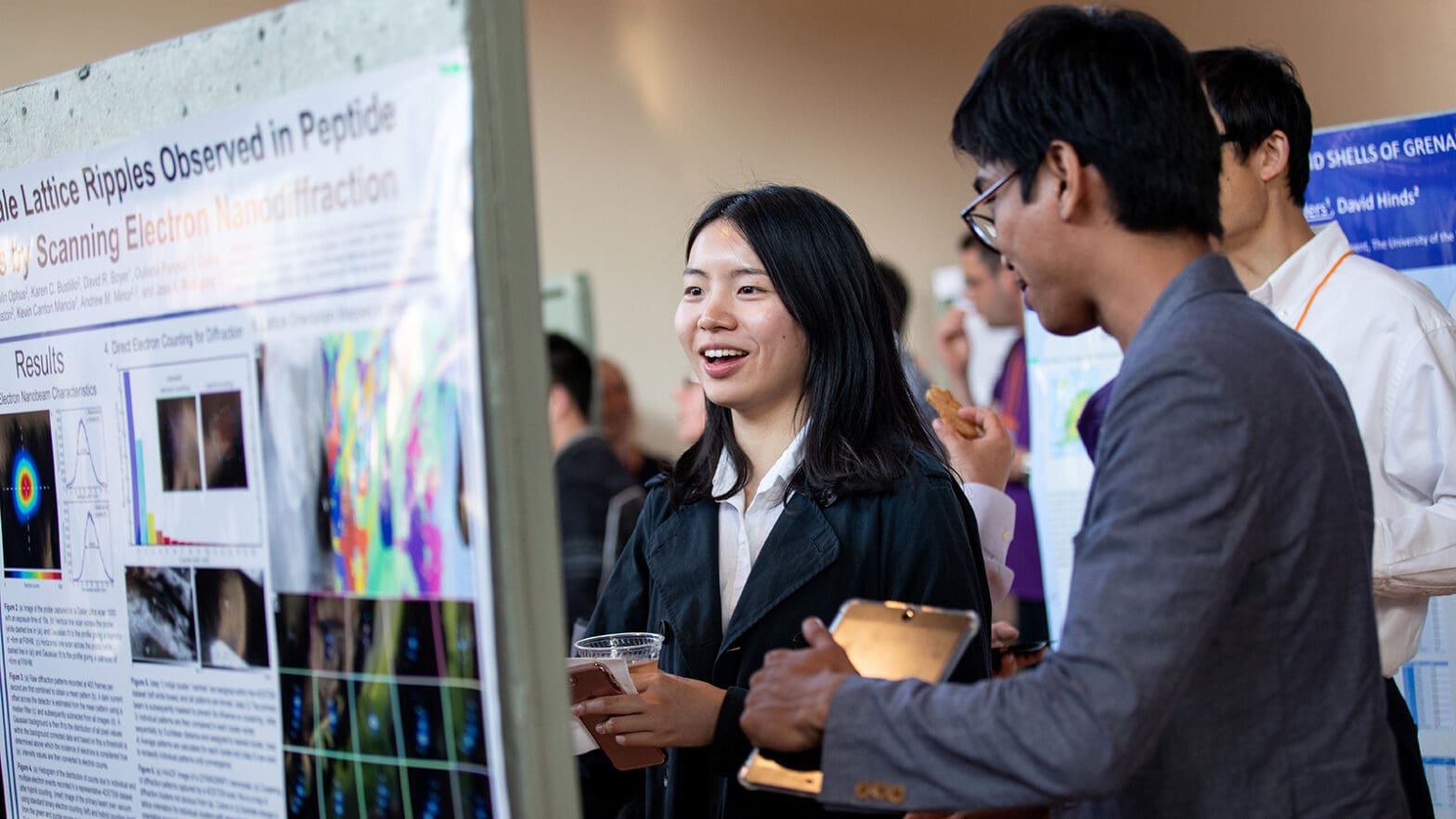
Nan Yao, director of PRISM’s Imaging and Analysis Center and lead-organizer of the conference, said that a common thread among the most cutting-edge techniques discussed at the conference is that they seek to maximize the information that can be extracted from the interaction of an electron and the sample that is being observed.
“A big takeaway from the conference was just how much information we can achieve from revolutionary capabilities of electron microscopy,” Yao said. This information falls in many areas, including structural, electromagnetic and chemical details: the ability to locate atomic and molecular positions in three dimensions; to determine the magnetic properties of atoms; and to identify the chemical contents of living cells.
Most attendees at the conference were not experts in all of these areas at once, “because we’re all used to the techniques we use and don’t always know what else is out there,” Yao said. “It was very valuable to have in the same room so many people who are pushing these boundaries in so many directions.”
")
One example of maximizing data from electrons is a new type of microscope known as the “Cryo-Electron Microscope” – an electron microscope that observes minute structures in ultra-cold liquids. The Krios Cryo-EM, which was recently installed in the Imaging and Analysis Center, is particularly useful for capturing the very short-lived chemical reactions inside living systems and high-performance materials.
This technique differs from conventional ideas of microscopy in that it is more about collecting and manipulating large amounts of data than viewing single images. “The new thing is we are talking about massive amounts of data, a massive number of images,” said Joachim Frank, a biophysicist at Columbia University who won the 2017 Nobel Prize for founding the field of single-particle cryo-electron microscopy. “If one thinks about electron microscopy one thinks about a picture. Now think about hundreds of thousands of images.”
")
“This whole technology is about bringing these data together into 3D visualizations,” Frank said. “This is something that is completely new.”
Another set of talks addressed an approach called scanning transmission electron microscopy (STEM), which combines the ability to focus on extremely small features with the ability to scan rapidly across wider areas. Princeton’s Imaging and Analysis Center recently installed an advanced instrument for this technique called a Titan Themis Double Spherical Aberration-Corrected STEM. This microscope is capable of showing feature as small as 0.06 nanometer, which is similar in size to a single atom of hydrogen.
As well as covering a range of techniques and topics of research, the conference brought together leading researchers from industry, national labs and academics from around the world, said Yao. “Electron microscopy is a tool that crosses disciplines and sectors. We are proud to offer a facility to bring people across these areas together to share knowledge and gain insights.”
Further background on the event, including the full program, is available at https://www.nature.com/natureconferences/fempl2018/index.html.